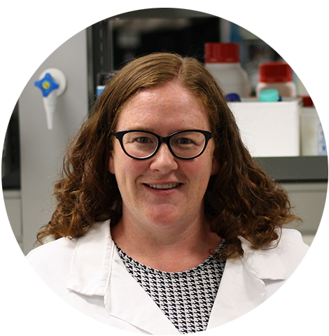
This past February Dr Juliette Cheyne participated in our Auckland Women in Science event. We took the time to get to know her a little bit better, and to have her share a wonderful research update about her current research on tracking activity in the brain in real time and how that relates to activity on behaviour or sensory input.
Why did you decide to become a researcher/ scientist/ clinician?
I enjoyed learning about the body and how it worked since I was very young as I grew up on a farm. During high school I had a really great science teacher and it was my favourite subject. I started University with the idea of doing medicine, but I wasn’t 100% sure. During 1st and 2nd year lectures by Sir Professor Richard Faull I was inspired to learn more about the brain. At the end of 3rd year I did a summer studentship in neuroscience and during those 10 weeks I became sure that I wanted to be a scientist as I really enjoyed being in the lab and working on a question that no-one knew the answer to.
What qualities do you think you need to have to be a researcher?
- Passion
- Resilience
- Dedication
- Good planning skills
- Working well as part of a team
- Trouble shooting
- Knowing when to stop and reassess the situation!
What inspires you to continue your work?
I hope that an increased understanding of the brain will help people in the future. My work is not directly translatable to help people right now, but I believe that every little bit helps. I enjoy working with a great team of people and inspiring the next generation of students - seeing their enthusiasm and progress is great positive feedback!
What advice would you offer young people looking to start a career in the science/research field?
Science is not always an easy path, so passion is very important as is good support - so find good mentors and ask lots of questions. Choosing the right lab for your Honours/Masters/ PhD/postdoc is really important. Make sure you find out about the inner workings of any lab before committing, talk to the lab members not just the principle investigator. Also check out the publication record of the lab and see if lab members came and left without publication. It’s also good to look into where previous lab members are now (e.g. are they still in science, do they have their own independent lab group etc). Don’t put your personal life on hold as there is never a perfect time to start a family in this career!
Research update
My research focusses on how brain cell activity controls behaviour and what happens when this goes awry in disease. My work utilises cellular recording in live mice (‘in vivo’) using a range of different imaging technologies. These techniques allow us to track activity in the brain in real time and relate the activity to behaviour or sensory input. All my research is performed with the support of my long-term mentor Associate Professor Johanna Montgomery.
To record brain cell activity, we genetically engineer brain areas of interest to make them flash when they become active. We then record movies of brain activity and synchronise them with videos of mouse behaviour or recordings of sensory stimuli presented to the animals. These types of experiments can be performed using different types of microscopes that each have their own strengths and weaknesses. One of the best types of specialist microscope for this type of work is not readily available in New Zealand. To overcome this problem, I teamed up with Associate Professor Frederique Vanholsbeeck and Professor Neil Broderick in the Physics Department at the University of Auckland. Our PhD student, Ashly Jose, is now customising our own two-photon microscope. This will be the first microscope in New Zealand to examine brain activity in live mice at high resolution, enabling activity in individual cells to be tracked.
During the past year Yukti Vyas, who will soon graduate her PhD, has established miniaturised microscope (‘miniscopes’) recordings in the lab while working as a Research Technician/Research Associate. These tiny (~3 grams!) microscopes can be used to record brain activity in freely moving rodents. In collaboration with Professor Cliff Abraham, from the University of Otago, we are applying this technology to better understand changes in the hippocampus in a mouse model of Alzheimer’s disease. Alzheimer’s disease is a progressive neurodegenerative disease that is the most common cause of dementia. Symptoms of Alzheimer’s disease include memory loss, disorientation, as well as mood and behaviour changes. These symptoms are caused by neuronal degeneration and cell loss that begins in the hippocampus, and spreads to the rest of the brain later in disease progression. We are imaging activity in hippocampal brain cells while mice perform memory tasks. These experiments will provide a real-time readout of the brain activity changes that underpin the hippocampal learning deficits observed in Alzheimer’s disease.
Another major aspect of my research is to understand more about hearing and auditory processing in mouse models of Autism Spectrum Disorder (ASD). ASDs are a set of developmental disorders defined by impaired learning, sensory disorders, communication difficulties, social deficits and stereotyped behaviours. The social and communication difficulties in ASD are thought to be due to the distorted processing of sounds, which in turn impairs language abilities. Masters student, Minchul Park, examined hearing in a mouse model of ASD by recording auditory brainstem responses from the scalp of mice in collaboration with Professor Peter Thorne. We found that these mice have normal hearing when they are young adults. However, we were curious as to whether there were any differences in sound processing higher up in the processing pathway, in the auditory cortex.
Throughout the auditory system, neurons are spatially organised according to which type of tones they prefer (high or low frequencies) - this is called tonotopic mapping. Masters student Maya Wilde examined whether sound responses are mapped differently in the auditory cortex of ASD mice. Our results show that there are differences in the representation of tones in the auditory cortex of ASD mice. Specifically, ASD mice showed an enlarged auditory cortex and more variability in tone frequency preferences. These findings indicate more poorly regulated cortical representation of sound in this model of ASD, which may link to the human phenotypes of auditory hypersensitivity and language impairments. Together with PhD student Pang Ying Cheung, we are currently examining the tonotopic maps in ASD mice with a higher level of detail. In future experiments PhD student, Ashly Jose, will examine these maps with single cell resolution using our customised two-photon microscope.
Lastly, together with PhD candidate Zahra Laouby, we have also begun applying these in vivo imaging techniques to rodent models of spinal cord injury in collaboration with Dr Simon O’Carroll. Following spinal cord injury, inflammation and neuronal damage occur and lead to loss of function. A treatment developed in Dr O’Carroll’s laboratory improves outcomes after injury but in order to fully understand how this treatment works it is important to be able to monitor cellular changes that occur in vivo. To do this Zahra will perform miniscope and two-photon imaging in the motor cortex and spinal cord.

Hear Dr Juliette Cheyne speak about her research at our upcoming event in Whangarei.


